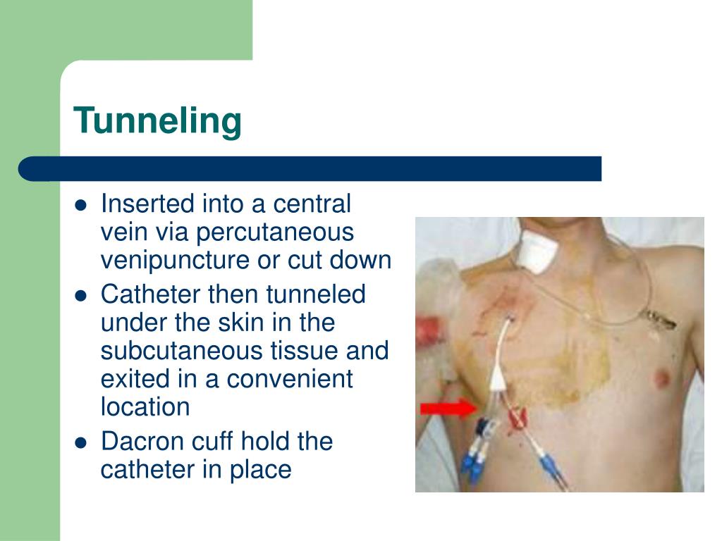
Step 8: Place needleless connectors on each catheter lumen. Maintaining control, gently advance catheter to premeasured internal length. Step 7: Slowly thread the catheter over the wire. Once dilator/sheath has been inserted, remove the dilator always maintaining control of the guide wire.
RIGHT SUBCLAVIAN TRIPLE LUMEN CATHETER SKIN
Using a circular twisting motion, gently slide the dilator/sheath assembly into the skin down to the depth of the vein, keeping control of the wire at all times.ĭepending on catheter size, you may need to dilate up to a larger wire. Step 6: Advance dilator/sheath assembly over wire. Perform skin nick with a straight scalpel blade. West J Emerg Med 10: 110-114.Step 5: If necessary, administer additional Lidocaine. Patrick SP, Tijunelis MA, Johnson S, Herbert ME (2009) Supraclavicular subclavian vein catheterization: The forgotten central line. Indian Journal of Anaesthesia 52: 337-339. Anesthesiology 95: 1377-1379.Ĭhauhan A (2008) Malpositioning of central venous catheter: Two case reports. StatPearls.Īmbesh SP, Pandey JC, Dubey PK (2001) Internal jugular vein occlusion test for rapid diagnosis of misplaced subclavian vein catheter into the internal jugular vein. But because of several anatomic advantage like larger diameter, relatively constant position, absent valves along with reduced risk of infection and thrombosis, it should not deter the clinician from using it as one of the sites for central venous access.ĭeere M, Singh A, Burns B (2022) Central venous access of the subclavian vein. ĭespite following recommended measures like lowering the shoulder, putting pressure over the supraclavicular fossa, using ultrasound for vein and guidewire locations, and observing for atrial ectopic, there is still a chance for catheter tip malposition. The causes may be a more horizontal orientation of the right and left brachiocephalic trunks, a longer insertion path (> 18 cm), and a changed orientation of the J-tip of the guidewire during the insertion.

The subclavian vein catheter is most commonly misplaced in the right internal jugular vein rather than the opposite subclavian vein. The likely cause for this misplacement may be that the guidewire in our case must have passed through the right subclavian vein, the right brachiocephalic vein, the left brachiocephalic vein, and finally the left subclavian vein.įigure 2: Chest X-ray showing correct placement of right internal jugular vein venous catheter. The catheter was removed, and the right internal jugular vein was correctly cannulated (Figure 2). The procedure was uneventful but the chest X-ray revealed the tip of the catheter to be present in the opposite subclavian vein (Figure 1). Because of difficult peripheral intravenous access, a central venous access was placed in the right subclavian vein under ultrasound guidance. After obtaining the informed written consent, we present an image of a 48-year-old female who presented to emergency with traumatic right above elbow amputation and blunt chest trauma. But mispositioning of the catheter tip into the contralateral subclavian vein is extremely rare. However, their mispositioning is not uncommon.

Its proper placement is necessary for long-term use.

Central venous catheter placement is associated with known risks and complications.


 0 kommentar(er)
